A life lost too soon.

On February 15 the world lost an amazing person all too soon. The passing of Kalena DeBrum has left us shocked and heartbroken. Kalena, a PhD student in our lab, was an internationally recognized scientist in the field of marine conservation.  Her fervent commitment towards preserving the health of coral reefs was matched only by her dedication to educating and empowering community members to be stewards of their waters, ensuring a legacy of sustainability for generations.

Through her influential role with the Marshall Islands Marine Resource Authority, and her dissertation research on reef fishes from the region, Kalena ascended to prominence as one of the world’s authorities on the coral reefs of the Marshall Islands.  Her careful dedication to working hand-in-hand with local community members, fishers and scientists represents the ethical benchmark for how environmental research should be conducted not only in Oceania, but globally.  At UW, she was an inspiration for the next generation of scientists, especially young Pacific Islander women.  

But Kalena was more than just a rising star in the world of marine conservation, and an exemplar for how community-based research should look, she was a force. Kalena was powerful. She took up space. She spoke truth to power. She was sharp as hell. Radiant. Hysterical. Goofy. Tenacious. Deeply inspiring. I promise we will not lose the momentum that you embodied, young lady. We will harness that inertia and carry your energy forward so that others can follow in the huge footsteps you leave behind.

We miss you, kiddo.

The Response of "Super Corals" to Extreme Climatic Environments

Coral reefs are essential habitats for marine life but are also the coastal ecosystem most impacted by climate change. Climate change is modifying the ocean’s environmental conditions through rising temperatures, acidity, and decreased dissolved oxygen, threatening the survival of coral reefs. Predicting how corals will react to upcoming climatic conditions is vital to forecast how these ecosystems might change in the future, but lab experiments are often limited by time, preventing long-term responses of corals to future conditions. This is why extreme reef environments, i.e. sites that already experience future-like conditions, are extremely valuable to investigate the response of corals to climate change. Such a site has been identified in 2017 in New Caledonia (lagoon of Bouraké, Southwest Pacific), and our current lab member Juliette had the opportunity to participate in a R/V field trip in 2020 with the goal of better understanding the metabolic specificities of corals thriving at this extreme site. 

Coral Incubatioin set-up

The lagoon of Bouraké is of particular interest because it is one of the only reef environments identified where the three climate stressors (T, pH, DO) predicted to impact corals co-occur.  However, despite these conditions, some coral species are healthy and give insight into how corals could persist in a changing ocean. To test whether the success of these “super corals” could be explained by changes in their metabolic rates, Juliette and Clément, supervised by Dr. Rodolfo-Metalpa (Institute of Research and Development, Nouméa) investigated the photosynthesis, respiration rates, and symbiotic characteristics of corals in the Bouraké lagoon and compared them with those of corals from a near-by control reef experiencing typical ocean conditions. 

Juliette on board the R/V, analyzing seawater conditions. 

Colonies from seven coral species were sampled at both the extreme and the control site and were incubated in conditions mimicking either present-day or future-like environmental conditions. For corals from both sites, and under both incubation conditions, photosynthesis, respiration and symbiont content were measured. This study allowed us to formulate several conclusions. First, symbiont density and chlorophyll content were as high or higher at the Bouraké site than at the control site. This is a demonstration of the good health of Bouraké corals, as corals tend to expel their symbionts when experiencing stress.  Second, the incubations revealed that no changes in the metabolism of corals occurred when exposed to short-term changes of environmental conditions. Lastly, measurement of metabolic rates evidenced significant differences in the respiration and photosynthesis of corals from the extreme and the control site. However, these changes differed among species, indicating that mechanisms underlying these shifts were species-specific rather than ubiquitous. The specificities of the observed responses of corals developing in extreme environments suggests that no unique response will determine corals tolerance to future conditions.

Coral sampling by Clément at the extreme site of Bouraké.

Jacquemont, J., Houlbrèque, F., Tanvet, C. et al. Long-term exposure to an extreme environment induces species-specific responses in corals’ photosynthesis and respiration rates. Mar Biol 169, 82 (2022).

For access to the full version of the published article, follow this link: https://rdcu.be/cOt8M


Invasive species on Caribbean deep reefs

Author: Sarah Panciroli
 
Invasive species pose a huge threat to marine biodiversity. One of the most notorious and alarming cases of this is the invasive lionfish that has rapidly invaded the tropical and subtropical western Atlantic coastal ecosystem since the late 1980’s. Voracious, predatory, and successful, this species owes its success to its morphological and behavioral traits. Lionfish have an unusual shape with venomous spines concealed in their feathery appendages protruding from their body. Along with this confusing shape, lionfish are outfitted in a maze of vibrant stripes and cryptic coloration. These defensive characters are accompanied by a wide range of feeding behaviors, a generalist diet, and protection from many parasites. This fish is rapidly decimating populations and becoming the locust of the sea.

Lionfish are typically found in coastal waters, where control over their invasion is in reach of scientists and management efforts such as incentivized lionfish derbies. This issue of massive invasion could be somewhat manageable if lionfish were exclusive to this shallow region, but in recent years they have been found in reefs deeper than 300 meters in the Caribbean region (Gress et al. 2017)— which means the devastating impact this creature has on ocean diversity may get a whole lot deeper.

Lionfish that occupy deep-reef ecosystems presents even greater concerns because shallow-reef environments possess resilient qualities, containing diverse populations and a large prey availability, mitigating effects of invasive predators. In contrast, deep-reef ecosystems have less habitat availability, limited species diversity and prey availability. Ecosystems like this can be easily exploited by an invasive species that can wipe out large volumes of biomass (Andradi-Brown et al 2017). Controlling lionfish populations is also an overwhelming feat, as derbies and other methods would be near impossible at depths 50-300m.

 Deep reef ecosystems have historically lacked the wealth of information that are available for shallow reefs, which has led to new scientific programs specifically looking to investigate biodiversity and conservation on deep reefs. The Smithsonian’s DROP (Deep Reef Observation Project) is one of these programs which has contributed to research on relationships between deep and shallow environments, focusing on biodiversity, conservation, and evolution. In 2017, UW partnered with DROP. Through this collaboration, the University of Washington pioneered a lionfish research project to learn more about the impacts of lionfish that occupied deep reef ecosystems. DROP uses submersibles that have the capability to collect lionfish occupying deep reefs, allowing us to ask whether lionfish on deep reefs occupy different trophic positions than lionfish on shallow reefs. The project was jump started when University of Washington graduate Megan Ewing was introduced to this question while working in the Burke Museum Fish Collection. 

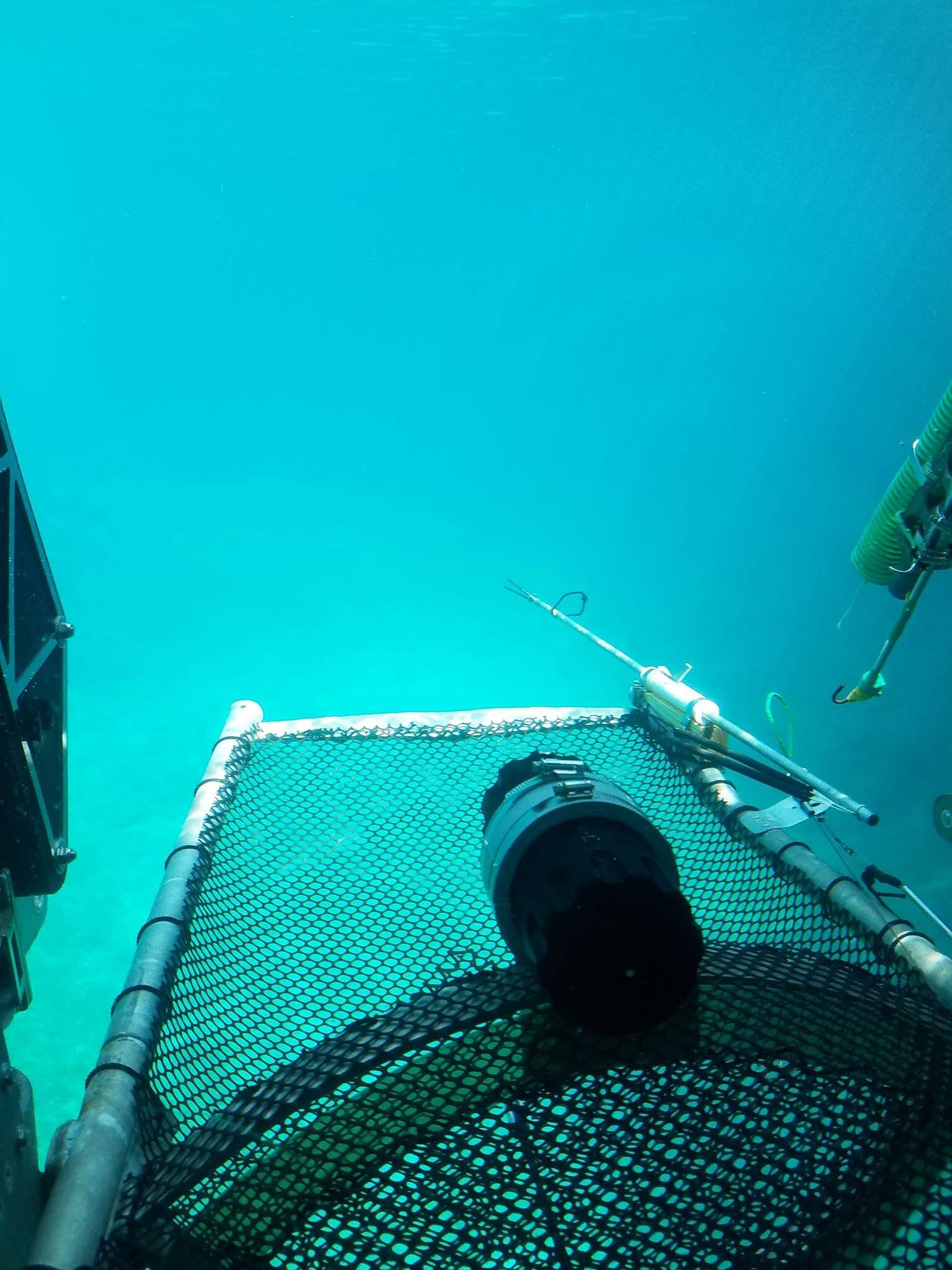
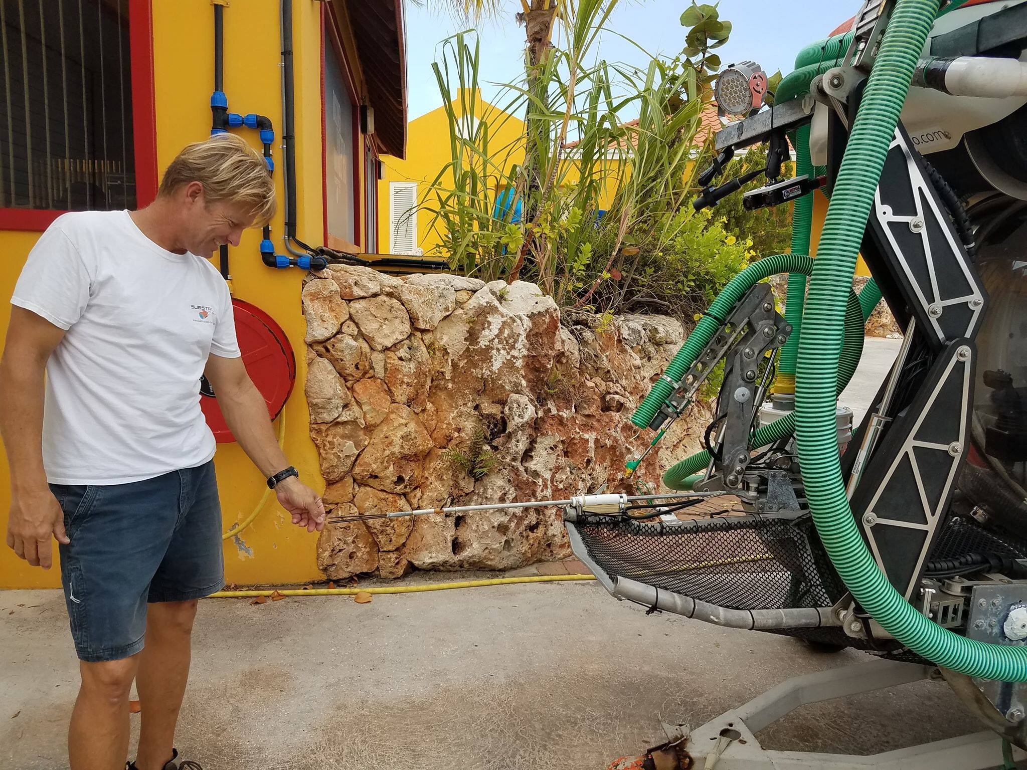
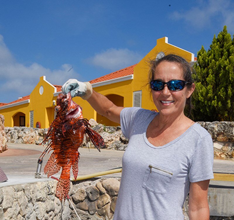
Trophic positions of deep-reef lionfish was a question that sparked the interest of undergraduate Megan Ewing in the Spring of 2020. While working in the fish collection, she was introduced to the numerous lionfish preserved in the labs deep freezer, collected by DROP using a submersible called the CURASUB in Curaçao. After several months of identifying and cataloging typically tiny specimens, Megan was excited to work with the larger creatures and answer questions related to the invasive lionfish. The basis of this question of diet and trophic positions relied on a concept called stable isotope analysis, using the two isotopes δ13C and δ15N. These elements would ultimately tell us two things: carbon isotope ratios would explain what environment the lionfish were eating, as carbon signatures found in a species can be matched to unique signatures found in specific marine habitats (deep reefs vs shallow reefs). Nitrogen isotope ratios show what trophic level an animal is eating, because nitrogen accumulates up the food chain. This background knowledge helped guide Megan’s hypotheses that deep populations of lionfish will have enriched δ15N values compared to shallow populations. This is because the increased energetic demands of more mature and larger fish found on deep reefs will lead to more prey consumption and a higher trophic position. Deep populations of lionfish will also have slightly depleted δ13C values compared to shallow populations as δ13C typically becomes depleted with depth (Fry et al. 1982, Fry et al. 1988).

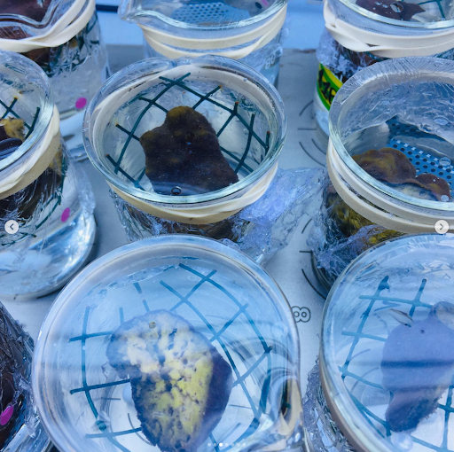
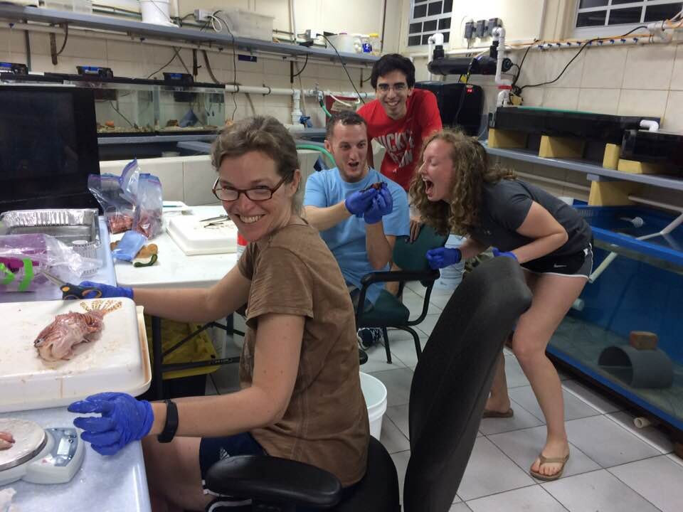
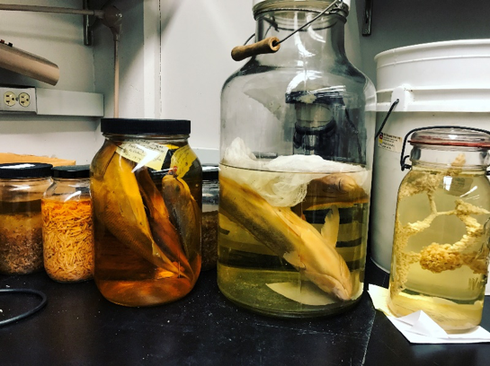
The lionfish project began with 76 fish captured off reefs in Curaçao, Caribbean, 31 collected in shallow reefs, and 45 from deep reefs. Megan extracted heart, muscle, and scale tissues to examine dietary changes over short, intermediate, and long time scales respectively. Once tissue samples were organized and extracted, they needed to be prepared for bulk stable carbon and nitrogen isotope sample, a process described through (Peterson & Fry 1987), which involved grinding and weighing each 3 tissue samples of all 76 lionfish. This may sound simple, but grinding each sample of lionfish alone took three months, with time consuming obstacles like extensive sterilization, assuring the samples were consistently a baking powder consistency, and funneling these samples into tiny vials. The next step of preparation involved weighing the small bits of powder produced from grinding. “Weighing” cannot simply capture the amount of effort exerted into this process. Each sample had to weigh between .4-.425 mg, a difference that is not detectable to the human eye and comparable to the weight of an eyebrow hair. A sample this small was transported using scoops the size of a grain of rice. Once the sample was finally in the correct weight range, it was folded in its vessel in a very particular shape and placed into a capsule on a 96 well plate, among many other samples. To call this process precise would be an understatement, and is a testimony to the amount of focus and determination that science sometimes requires. Adding to the intensity of this work, a large portion of it was done in Megan’s home during a global pandemic. When everything was finally in its perfect place and measured state, the samples were sent to the UC Davis stable isotope facility to determine the δ13C and δ15N ratios.

Proceeding the lengthy scientific methods, the highly anticipated results were eventually received from the UC Davis isotope sampling lab in 2021. For all tissues, there was no significant difference in δ 13C between shallow and deep populations, but the δ 13C range of deep populations is broader than and overlapping with shallow populations. However, deep population lionfish had enriched δ 15N when compared to the shallow reef lionfish. In addition to confirming Megan’s original hypothesis, this information presented several conclusions.  The increased and overlapping range of δ13C for deep populations when compared to shallow populations indicates that not only do they likely have larger habitat range when compared to shallow populations, but there could also be a vertical migration by the deep reef populations to shallow environments (Fry et al. 1982). The increased δ 15N in deep reef lionfish also indicates that they occupy a higher trophic position, as nitrogen accumulates up the food chain.

The implications of these results provide areas of concern, but also room for hope. If deep reef lionfish are indeed undergoing vertical migration, controls on these populations would be more feasible with greater management pressure exerted when these populations are feeding in shallow reefs.  However, the higher trophic position of deep reef populations has major consequences for their surrounding habitats. Lionfish are invasive generalist predators, so as their trophic position increases, abundance and diversity of native prey will drastically decrease. Increased studies on deep reef populations to truly understand the implications of higher trophic level lionfish feeding will most likely proceed this work and strengthen this study. Research could be done to determine when these populations are migrating to shallow environments, to make management efforts more impactful to eradicate this population. Ultimately, there a distinction to be made between these two populations of lionfish- providing greater motivation in exterminating this invasive species when it threatens a less researched habitat in the ocean that is clearly more vulnerable to the effects of a high trophic feeding animal.


Behind the researcher:

The extensive research and scientific experimentation required to answer this question would not be possible without University of Washington’s graduate Megan Ewing. Many factors contributed to Megan gaining interest on marine related projects such as these. For Megan, the college experience was novel in her family, being the first of her immediate family to go to a university. This situation added several challenges, such as a lack of guidance and support when applying to college, and figuring out the intricacies and confusion that are associated with applications. Nevertheless, Megan was successfully accepted to the University of Washington, a highly acclaimed and competitive college. The first years of college are met with curiosity and typically an uncertainty in academic direction. Megan wasn’t originally drawn to marine and fishery studies, and instead was dead-set on business, specifically marketing for nonprofits and sustainable ocean companies, but when the major didn’t work out she needed a plan B. Keeping the passion for the ocean and sustainability in mind, Megan then went to the marine biology advisor who recommended taking Fish 311, a course taught by Luke Tornabene that encompassed fish biology and evolution. The first lab included a tour of the UW fish collection, a vast room containing millions of preserved specimen and endless opportunities for research: Megan was sold. Marine Biology satisfied the passion of conservation and knowledge of the ocean, but also contained hard core sciences that wasn’t originally anticipated, adding to the appeal. These qualities made marine biology turn from a childhood dream as it is often regarded, to a serious career which Megan was determined to pursue. Once settled in the major, Megan reached out to Luke to volunteer in the fish collection, where she worked on fish identification, collection organization and was interested in ecology, reproduction, and behavior predation. After countless hours working with tiny specimens, Megan was introduced to the onslaught of lionfish collected from Curaçao. Given her interest in ecology and feeding behaviors, Megan took on the lionfish project, eager to investigate important topics of invasive species and work with larger specimen. Megan continues to write up the research paper on her investigation and is currently employed as an education volunteer coordinator at the Friends of Issaquah Salmon Hatchery (FISH). Megan explains this is a “non-profit with the mission of protecting our salmon and ensuring their survival for future generations through education, advocacy, and outreach." As an education/volunteer coordinator, she works with local schools to teach children (K-12) about salmon life cycles, salmon habitats, ecology, watersheds, and the role of hatcheries.

As for the future, Megan plans to go back to school for her PhD, ideally to study reproduction in fish, and continue to find research projects that involve field work and laboratory methods. Her end goal is to become an educator, such as becoming a professor at a research heavy university just like her undergraduate alma mater.

The work provided by Megan was highly valuable and a fantastic undergraduate achievement that provided evidence that can help answer questions about this hot topic in conservation. We are positive she will continue to explore the ocean and share her knowledge with others, positively contributing to the scientific community.


Innovation Award and NSF Scanning Grant Pave Way for New Research


  Normal
  0


  false
  false
  false
  
  EN-US
  JA
  X-NONE


&n…

Preserved adult and juvenile fishes from the UW Ichthyology Collection, photo by Katlyn Fuentes

It’s an exciting time for the Fish Systematics and Biodiversity Lab! Two recent awards will be helping to pave a way for new, cutting-edge research involving our very own UW Fish Collection.

Established in 2014, the UW Innovation Awards have helped fund a total of 16 faculty projects with an approximate $3 million supporting ground-breaking research in areas such as education, health care, and now fisheries. A joint project proposed by Dr. Chelsea Wood – a parasite ecologist and fellow Assistant Professor at the UW School of Aquatic and Fishery Sciences – and Dr. Luke Tornabene, was selected to receive $296,067 over the next two years to understand long-term change in parasitism of Pacific Northwest fishes. The proposed project will use specimens from the UW Ichthyology Collection for parasitological dissections – a method pioneered in Wood’s lab.


To help fill the gap in historical records of marine outbreaks, parasite assemblages will be reconstructed for 10 species of Puget Sound fishes from 1880 to the present day, with the species identification and number of parasites found on these specimens recorded.

Why might this be important to study, you ask?

Though outbreaks of marine diseases have become a regular occurrence, the lack of historical baseline data of these mass-mortality events have left scientists at a loss for assessing the frequency or severity of these events. By generating baseline data, Wood and Tornabene hope to determine whether marine epidemics are increasing in frequency and magnitude, and therefore predict not only the time but the location in which these events might occur in the future.

(Among the joint-project proposed by Dr. Tornabene and Dr. Wood, there were four other recipients of this year’s awards, with projects focusing on studying glacial retreats in the western U.S. and the effects of Arctic sea-ice in nutrient content of algae in polar environments.)


  Normal
  0


  false
  false
  false
  
  EN-US
  JA
  X-NONE


&n…

A CT scan of a spotfin hatchetfish, Thoracocharax stellatus; photo by Adam Summers – University of Washington, Friday Harbor Laboratories

oVert GRANT

In addition to the UW Innovation Award, a recent $2.5 million grant from the National Science Foundation will help to fund a project of which will shape zoological-specimen-based research for generations to come. Over the next four years, UW and 15 other institutions from around the world will CT scan an approximate 20,000 museum specimens in hopes of creating a digital encyclopedia of all known extant vertebrates. As one of the largest fish collections in the world, the UW Ichthyology Collection will be making considerable contributions to this project by partnering with researchers at Friday Harbor Laboratories (in San Juan Island, WA) to #ScanAllFishes. After digitizing these museum specimens, those involved will then publish the scans for anyone who might wish to view them – including other researchers, students, and the public!

FUTURE IMPLICATIONS

The implications of these two awards are astronomical. The products from these research projects will go on to benefit the masses by allowing public-access to never-before-seen research and helping predict marine epidemics which have the potential to impact multiple facets of every-day life – such as public health and ecosystem services.

Our natural history museums act as bridges between the scientific and public communities! By continuing to fund projects that involve the use of specimens from natural history museums, we’re proving that these collections are worth more than the occasional trip to a museum on a rainy day. These specimens can be utilized for far more than just systematic-based projects… perhaps even projects that have the potential to change the field of environmental science as we know it.

So congratulations to all those involved in these two ground-breaking research projects! Make sure to check back frequently for future updates as we progress in our research!

-Katlyn Fuentes


The lab heads to Rochester NY to showcase latest research!

From left: Luke, Jenny, Katherine, Calder, Dara, Emily

From left: Luke, Jenny, Katherine, Calder, Dara, Emily

For the Fish Systematics and Biodiversity Lab, summer means time for fieldwork, workshops, and conferences. In August, six members of the lab flew to Rochester NY to present at the Joint Meeting of Ichthyologists and Herpetologists (JMIH). For five days we were able to share all of our hard work, discuss plans, and most importantly, have fun with colleagues past, present, and future.


Katherine

Katherine

Katherine Maslenikov (collection manager) helped kick-off the event by leading a half-day workshop teaching best practices when caring for natural history collections – an honor merited by her years of hard work with the University of Washington’s Ichthyology Collection. Over the following days, Katherine, Luke, and four of his students presented on the various research projects happening in the lab. Katherine co-authored a poster looking at ontogenetic series of fishes through contrast-enhanced CT scanning. 


Jenny

Jenny

M.S. student Jenny Gardner shared her exploration in CT scanning snailfishes in the family Liparidae, who get their claim-to-fame by being the deepest living vertebrate on earth! She is using the 3D scans to characterize the internal morphology, particularly of soft tissue, in these fishes and how it differs between species. CT scans are traditionally used for detecting hard tissue such as bone, but Jenny is experimenting with making soft-tissues more visible by staining her fishes using iodine.


Calder

Calder

Calder Atta, another M.S. student, presented his field surveys from a previously unsampled coral reef in the Red Sea, a poorly studied region to begin with. His work shows that there is a diverse community of cryptobenthic fishes and provides evidence for a unique composition of reef fishes in the area, potentially including some new species. He also highlights the importance of using both visual transects and cryptobenthic collections in biodiversity surveys.


Dara

Dara

Dara Yiu recently graduated and shared her completed undergraduate thesis on the dwarf goby Eviota. Her work followed the discovery of two different color morphs within the species Eviota atriventris. She used genetic markers to test if the color morphs represent distinct lineages within the species and found that the gobies may be in the process of splitting into separate species.


Emily

Emily

Both Luke and Emily McFarland (rising junior undergrad) presented on fish communities that live on Caribbean deep reefs. Emily focused on how different trophic guilds (Carnivore, Herbivore, Planktivore, etc.) are distributed through depth and between sites, as well as the time-consuming work of extracting data from submersible dive footage. Luke brought attention to a distinct community of fishes that inhabit these deep zones that have been neglected by scientists for years, now called “rariphotic” reefs.


Rochester Convention Center, on the last evening of the trip. 

Rochester Convention Center, on the last evening of the trip. 

The 2018 JMIH was a great opportunity for students to network, and for our lab to connect with fish biologists around the world. Without these connections, we would not be able to study the wonderful world of fishes!

-Calder Atta

Girl Power!! The Ladies of the Fish Systematics and Biodiversity Lab Receive Funding for their Research

The start of 2018 has been busy, but also productive, for the members of the Fish Systematics and Biodiversity Lab. While Luke enjoyed teaching Biology of Fishes solo for the first time,  his students cashed-in on some big grants and scholarships. The following is what lab members Dara Yui (senior undergrad), Marta Gómez-Buckley (Ph.D. student), and myself, Sarah Yerrace (junior undergrad) have spent the last several months working on.

Dara received the Mary Gates Research Scholarship in the fall of 2017. She was awarded $5,000 to support her genetic research on the dwarfgoby genus Eviota. Thanks to wide-spread use of DNA sequencing and molecular phylogenetic analyses in coral-reef fish taxonomy over the last decade, the description of new Eviotaspecies has exploded in recent years. The genus now comprises 113 species - an almost 500% increase from the 19 species that had been described before the 1970’s. Dara will be focusing on the Eviota atriventrisspecies complexPreliminary analyses show that within this species there are two different lineages showing subtly different color morphs from Cenderawasih Bay (Indonesia) and Milne Bay (Papua New Guinea). To clarify the taxonomy of the two lineages, Dara has been sequencing mitochondrial and nuclear genes for all available specimens as well as examining morphology. Her research will provide insights on the mechanisms of speciation in this rapidly diverging group of fishes. 

Eviota atriventris, photo by Jack Randall. 

Eviota atriventris, photo by Jack Randall. 

Marta received $6000 from the Hall Conservation Genetics Research Fund. As the name suggests, this fund is specifically for graduate students working on conservation genetics. Combining systematics, phylogenetics, conservation genetics, and community ecology, Marta will be evaluating the dynamics of coral reef ecosystems. She will do this by focusing on cryptobenthic reef fishes, as they contribute disproportionately to the overall diversity and energy transfer on a reef. 

Marta will be comparing environmental DNA (eDNA) and cryptobenthic reef-fish surveys to see if eDNA can accurately inform us about the diversity and community composition of cryptobenthic fishes. If eDNA can be used to monitor ecologically sensitive cryptobenthic communities, it would be a good indicator for reef degradation and habitat loss. This method could be more cost- and time-effective than surveying populations through destructive sampling.

Cryptobenthic reef fishes of Tonga, collected and photographed by Marta Gómez-Buckley.

Cryptobenthic reef fishes of Tonga, collected and photographed by Marta Gómez-Buckley.

Like Dara, I also received $5,000 from the Mary Gates Endowment. My research is also focusing on a small goby, Risor ruber. The common name for this fish, the tusked goby, comes from the two to four outward-facing canines on the upper and lower jaws of the fish. Preliminary genetic data indicates that there are eight different genetic lineages and ecological data shows that these lineages have species-specific commensal relationships with different sponge hosts. Host specialization could be the mechanism for the reproductive isolation causing genetic divergence, and ultimately speciation. I will be using a combination of fresh and preserved photos, cleared and stained specimens, and CT scans to analyze geometric morphometrics and meristic differences between lineages, and ultimately determine whether there are morphological differences that correspond to the genetic and ecological data within this group. 

Risor ruber, the Tusked Goby.  Photo by Carole C. Baldwin. 

Risor ruber, the Tusked Goby.  Photo by Carole C. Baldwin. 

Finally, in other Girl-Power related news, the Fish Systematics and Biodiversity lab was part of another female-led collaboration that also recently received research funding. The Deep Reef Observation Project, or DROP, is a program that is leading the way in exploring deep-reefs of the Caribbean. Carole Baldwin, Curator of Fishes at the Smithsonian National Museum of Natural History and Director of DROP, teamed up with Luke and Ross Robertson (Smithsonian Tropical Research Institute) to acquire funding to help establish a new base of operations for DROP in Roatan, Honduras.  The team will conduct 20 submersible dives on the Idabelmanned submersible over the next two years, collecting samples that will help us understand the evolution and connectivity of deep-reef fish communities.

Idabel submersible, located at Roatan, Honduras. 

Idabel submersible, located at Roatan, Honduras. 

Stay tuned for more updates from the members of the Fish Systematics and Biodiversity lab! 2018 is shaping up to be a big year full of discoveries.

UW Fish Collection shows off its selection of Halloween Horrors

By Emily McFarland
When October comes through and the sky gets dark and the fog rolls in, the time comes to celebrate the creepy crawler critters that lurk in the shadows. That’s just what we did on Friday, October 20th, at the Burke Museum for an event called Creepy Crawly Cocktails. Biologists of all sorts selected the creepiest, crawliest specimen from our collection to put on display for the public, and I was lucky enough to participate in showcasing some terrifically terrifying individuals from the fish collection. As much as ichthyologists may love fishes, nobody can deny that some true terrors lurk in the depths—and sometimes even in the shallows!


     We gathered an impressive array of horrors of all sorts, from the bizarre, like the oozing hagfish and strangely modified ratfish, to the downright fearsome, like the gnarly teeth of the lancetfish and alien-esque jaws of the moray eel. We even presented the handsome lionfish. Although its quills and toxins may be nasty, the true horror of the lionfish is more of an environmental sort—these voracious hunters are an invasive species in the Caribbean, and with no natural predators, they’re impacting the natural biodiversity of the area before we can even discover it! A true nightmare for ichthyologists everywhere.


     The event was a grand success! Loaded with questions, participants flocked to our table. They marveled at the bizarre reproductive strategy of the ratfish, clad with not one, not two, but four sex organs, one of which being located on the forehead. They carefully examined a bottle of wine, confiscated by the U.S. Fish and Wildlife Services for its unusual ingredient profile: ginseng, goji berries, shaving of elk and antlers, and most shockingly of all, whole seahorses. They recoiled with disgust and swore off swimming at the sight of the jawless, many-toothed mouth of the lamprey. Most importantly, they got the opportunity to engage with our underwater world and catch a glimpse of just how incredible—though occasionally frightening—it can be.


  Normal
  0


  false
  false
  false
  
  EN-US
  X-NONE
  X-NONE


  …

Pictured: Our bone-chilling display

burke2.jpg
burke4.jpg
burke5.jpg

GIRLS IN SCIENCE! Katherine, Sarah and Jalene train the next generation of women ichthyologists!

Story by Sarah Yerrace

For two hours every Monday, from April 17th to May 22nd, the UW Ichthyology Collection was busy with 16 girls, each working on describing a ‘new’ species. The girls —all from different Seattle high schools and all part of the Burke Museum's Girls In Science program — split into small groups to not only measure and count features on their ‘new’ species (Hexagrammos stelleri, Icelinus filamentosus, Artedius lateralis, Xeneretmus leiops, or Pholis clemensi) but also compare these measurements and counts from two other species within the same genus. Jessica Heide, a scientific illustrator, helped the groups draw a key morphological feature that varied between the three species. The Icelinus group, for example, chose to illustrate the unique dorsal fins of their three species while the Pholis group decided to draw the pigment patterns on the body of their fishes. By the end of the six weeks, the groups produced a poster, complete with a full write-up from the introduction to the acknowledgements.

UW Fish Collection staff members and part of the Girls in Science training team, Jalene Weatherholt and Sarah Yerrace.

UW Fish Collection staff members and part of the Girls in Science training team, Jalene Weatherholt and Sarah Yerrace.

The first session started off with a tour of the collection and an introduction to basic fish anatomy. The girls got their hands on some preserved specimens and practiced using keys to identify the fishes down to the family. While they may have been hesitant at first, none of the girls were squeamish around the preserved fishes and things picked up by the second session. They readily looked into a gaping mouth of a gadid to see its pharyngeal teeth and gill rakers. The teams worked great together to get the measurements done efficiently with every member assigned to a specific set of measurements for consistency across the three fishes. When it came time to create the poster, some of the teams got creative and themed their posters around a personality they saw in their fish. The Xeneretmus group, for example, saw poachers as a dragon like creature. They drew dragons on their poster and used an old script font. The new Latin name for their fish was Xeneretmus draco. While the format of the posters may not have been traditional to science, the girl’s enthusiasm was encouraging to see. At the end of the six weeks, the girls received some pins with fishes on them to remind them of the experience. Some of the groups even exchanged contact information to stay in touch. A mother said she has never seen her daughter more outgoing and comfortable around others. She said that when her daughter saw other women doing science, she felt like she could do it too. That is ultimately the goal of the Girls in Science program: to inspire young women and increase accessibility to STEM fields. 


Expedition to St. Eustatius

Last month our lab teamed up with with Smithsonian for the first-ever submersible exploration off St. Eustatius, one of six islands in the Dutch Caribbean.  St. Eustatius, or "Statia" as its known to the locals, is a tiny island with just a few thousand permanent residents in the Eastern Caribbean.

Upon arrival in Statia on April 14th, 2017 the DROP team boarded the R/V Chapman – a 127’ research vessel and mothership to the manned submersible Curasub (Substation Curacao). The goal of this trip was to explore deep reefs between 50 and 300 m and, for the first time, describe the communities that live at these depths off St. Eustatius. Reefs and other hard-bottom areas between 50 and 300 m are too deep for conventional scuba diving and are rarely surveyed by deep-diving submersibles or ROVS, making this diverse zone an area of the ocean that science has largely missed.  Using the Curasub, our team made five dives off the west coast of Statia, recorded more than 2000 visual observations of fishes and their depths of occurrence, and collected more than 350 samples of deep-reef fishes and invertebrates. Highlights from this trip are remarkable—at least eight species of fishes that are new to science and have yet to be formally described! These include two species of gobies (small, bottom-dwelling reef fishes) that are currently known only from Statia.  A total of 38 species observed on our dives have never before been recorded from St. Eustatius—a number that is even more impressive considering that the most comprehensive review of the fishes of St. Eustatius was just published in 2016. This means that the plethora of new records from this trip is not simply due to scarcity of published data from St. Eustatius, but instead, highlights the importance of sampling from hard-to-reach habitats like deep reefs.

In addition to finding new species or new records from St. Eustatius, the DROP team is also interested in finding out which deep-reef species are widely distributed and common in the Caribbean.  For example, deep-reefs off Statia were home to large numbers of Pugnose Bass (Bullisichthys caribbaeus), Sabre Goby (Antilligobius nikkiae) and Bicolor Basslet (Lipogramma klayi). These three species have also been recorded in large numbers from deep reefs off Curacao, Bonaire, Dominica, and Roatan Island. By collecting DNA samples of these species from each locality, DROP researchers can use cutting-edge population genetic analyses to begin to learn how deep reefs are connected across long distances, and ultimately understand whether distant localities can serve as a ‘refuge’ for local populations that experience declines due to habitat degradation or invasive lionfish.

The DROP team also measured water temperatures along the deep-reef slope in an attempt to understand whether temperature is correlated with how deep fishes live on the reef slope. Preliminary data suggest that fishes living in the warm water off St. Eustatius occupied deeper depths than they did off the cooler waters of Curacao, Bonaire and Roatan. This may mean that fishes are indeed choosing their preferred depths based on temperature differences. The implications of this finding are paramount, as sea-surface temperatures are projected to rise in response to climate change, and fish may seek these poorly-studied deep-reef habitats in search of cooler temperatures in the future.  Finally, a smaller DROP team remained on board to sample small to tiny fishes and invertebrates that inhabit sponges, more than 60 of which were brought to the surface by the Sirenas research team during five days of sub diving to depths of 50-250 m. Among the highlights of this collection were a weird shrimp, possibly a shell-less snail called a nudibranch, and one yellow organism with a soft exterior and hard interior that scientists couldn’t identify at all!

We are grateful to the crew of Substation Curacao and the R/V Chapman for this amazing opportunity. Sub diving in choppy seas such as those encountered in Statia is challenging, and it requires talented and dedicated teams in the sub, on the surface boat, and on the ship. As owner of Substation Curacao and the R/V Chapman, Adrian “Dutch” Schrier, is fond of saying, “if this were easy, everybody would be doing it.” It is not easy. Thank you!

Pictures up from Behind the Scenes Night at the Burke Museum

Last month the Burke Museum held a special Behind the Scenes night for members of the museum. The even was well attended, with several hundred guests touring the ethnology, biology and paleontology collections.  The Fish Collection was well represented, showcasing the Five Facets of Familiar Fishes at Burke (cleared/stained collection, alcohol specimens, otoliths, skeletons, and eggs/larvae). Videos of our recent submersible expeditions were also featured on the large projector in the Burke Room, with specimens of representative Caribbean deep-reef fauna for visitors to see first hand.  Students Sarah Yerrace, Rachel Manning, Sam Ghods, and their friends/family came out to help Katherine and Luke showcase the collection and educate the public on the importance of natural history collections.  Overall the night was an incredible success, and we cannot wait to do it again next year!